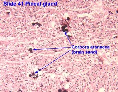
Corpora arenacea
| Brain sand | |
|---|---|
| Details | |
| Identifiers | |
| Latin | Corpora arenacea |
| Code | TH H3.08.02.3.00007 |
Corpora arenacea (or brain sand) are calcified structures in the pineal gland and other areas of the brain such as the choroid plexus. Older organisms have numerous corpora arenacea, whose function, if any, is unknown. Concentrations of "brain sand" increase with age, so the pineal gland becomes increasingly visible on X-rays over time, usually by the third or fourth decade. They are sometimes used as anatomical landmarks in radiological examinations.
Chemical analysis shows that they are composed of calcium phosphate, calcium carbonate, magnesium phosphate, and ammonium phosphate.[1] Recently, calcite deposits have been described as well.[2]
External links
Vígh, B; Szél, A; Debreceni, K; Fejér, Z; Manzano e Silva, M. J.; Vígh-Teichmann, I (1998). "Comparative histology of pineal calcification". Histology and histopathology 13 (3): 851–70. PMID 9690142.
References
- ↑ Bocchi, Giancarlo; Valdre, Giovanni; Valdre, Giovanni (1993). "Physical, chemical, and mineralogical characterization of carbonate-hydroxyapatite concretions of the human pineal gland". Journal of Inorganic Biochemistry 49 (3): 209–20. doi:10.1016/0162-0134(93)80006-U. PMID 8381851.
- ↑ Baconnier, Simon; Lang, Sidney B.; Polomska, Maria; Hilczer, Bozena; Berkovic, Garry; Meshulam, Guilia (2002). "Calcite microcrystals in the pineal gland of the human brain: First physical and chemical studies". Bioelectromagnetics 23 (7): 488–95. doi:10.1002/bem.10053. PMID 12224052.
External links
- Histology image: 14401loa – Histology Learning System at Boston University
- Histology image: 41_03 at the University of Oklahoma Health Sciences Center - "Pineal gland"
- Garma-Aviña, A. (2000). "Excretory Plugs from the Choroid Plexus in the Cerebrospinal Fluid of Dogs with Neurological Disease: Possible Role in the Formation of Corpora Arenacea". Journal of Comparative Pathology 123 (2–3): 146–51. doi:10.1053/jcpa.2000.0405. PMID 11032668.
| ||||||||||||||||||||||||||||||||||||||||||||||